
ACL reconstructive surgery is the only way to effectively treat a torn ACL. During surgery, a graft is taken from a chosen tendon and threaded through a hole drilled in the tibia into the femur. The entire process lasts approximately two hours.
After surgery, patients must wear an ACL brace and go through six to nine months of physical therapy. During that period, patients have to do strengthening exercises such as leg press and squats. Most athletes are able to recover completely after surgery.
After surgery, patients must wear an ACL brace and go through six to nine months of physical therapy. During that period, patients have to do strengthening exercises such as leg press and squats. Most athletes are able to recover completely after surgery.
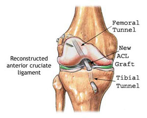
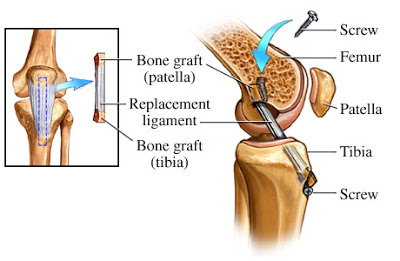
ACL reconstruction surgery uses a graft to replace the ligament. The most common grafts are autografts using part of your bodies, such as the tendon of the kneecap (patellar tendon) or one of the hamstring tendons. Another choice is allograft tissue, which is taken from a deceased donor.
Repair surgery is usually used primarily for cases where there is an avulsion fracture; this is separate of the ligament and a piece of the bone from the rest of the bone. In this case, the bone fragment t connected to the ACK is reattached to the bone.
ACL surgery is usually done by making small incisions in the knee and inserting instruments for surgery through theses incisions (arthroscopic surgery) In several cases; it is estimated by cutting a large incision in the knee during open surgery.
What Happens
During the Surgery ?
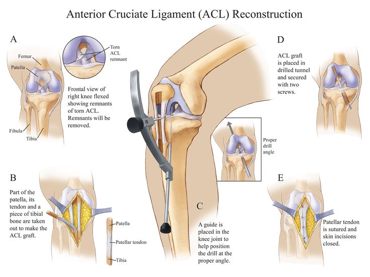
During Anthropic ACL reconstruction, your surgeon will make several small incisions- around two or three near the knee. The sterile saline(salt) solution will be pumped into the knee through one of the incisions to expand it and wash the blood from the area. This will allow the surgeon to see the structure of the knee more easily.
The doctor will insert an Arthroscope within one of the other incisions. A camera at the end of the arthroscope sends images from the inside of the knee to a Visual monitor in the operation room for the doctor to see.,
Surgical drills will be placed through other small incisions. The doctor drills small holes into the lower and upper leg bones where the bones are close to either a the knee joint. The holes will be for tunnels in which the graft will be anchored.
Another incision is made within the knee to take the replacement tissue or graft.
The graft is removed from the tendon at the front of the knee below the patella tendon or kneecap; which includes tow small pieces of bone called bone blocks on the ends of the time. One piece of bone is removed from the knee cap, and the other is removed from a part of the lower leg bone near the knee joint. This type of graft will allow the patient to heal better due the tendon being attached to its original bone, and the pieces of the bone just need to heal into their new placement.
If the autograft comes from the hamstring, bone blocks are removed. This type of graft can allow the knee to look more normal after it heals, due to the tendon from the front of the knees not being used. It may be easier to add additional tissue from a deceased donor called, an allograft to this particular type of graft.
ACL Reconstruction with Hamstring
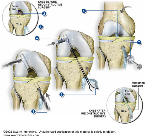
This procedure replaces a damaged or torn anterior cruciate ligament (ACL) with a portion of hamstring tendon from the patient’s leg. The ACL connects the front top of the tibia (lower leg bone) to the rear bottom of the femur (thigh bone). The hamstring tendons attach the hamstring muscles to the lower leg.
The excess autografts are trimmed away. The new parts of the knee are then tested by flexing and extending the knee through its full range of motion.
- Autograph Prepared
Through a small incision below the knee, portions of the knee, portions of the hamstring’s semitendinous and gracilis tendons are separated from the muscle but left connected to the tibia. These strips are braided together to create a section of tendon called an autograph, which will be used to replace the damaged ACL. - Torn ACL Removed
The rest of the procedure is preformed through small incisions on the sides of the knee. The surgeon uses a small video cameral called an arthroscope to see inside the knee during a procedure. With the knee flexed, the damaged ACL is cleared away.
Guide Pin Inserted A pin is inserted diagonally, from the tibia to the femur. The surgeon will use the pin as a guide to recreate the ACL. - Tunnel Created
The surgeon follows the guide pin, drilling a tunnel through the tibia and femur. - Horizontal Screw Inserted
A second tunnel is drilled to intersect with the femoral tunnel, and a horizontal screw is partially inserted. The hamstring grafts will be looped over this screw. - Graft Strand Secured
In order to pull the hamstring over the horizontal screw, a graft-passing strand is captured by the bone mulch screw within the femoral tunnel. The horizontal screw is then advanced and embedded into the bone securely. - New ACL Created
The hamstring grafts are tied to one end of the strand and pulled up through the joint and over the horizontal screw to create the new ACL. - Knee Straightened
After the knee is straightened, the loose ends of the grafts are pulled tight and held securely to the tibia bone with a washer that has nail-like spikes and a screw.
The excess autografts are trimmed away. The new parts of the knee are then tested by flexing and extending the knee through its full range of motion.
Recommended reading :
http://orthomedctr.com/anterior-cruciate-ligament-reconstruction.php
http://orthomedctr.com/anterior-cruciate-ligament-reconstruction.php
